
How a corneal scar changes your vision
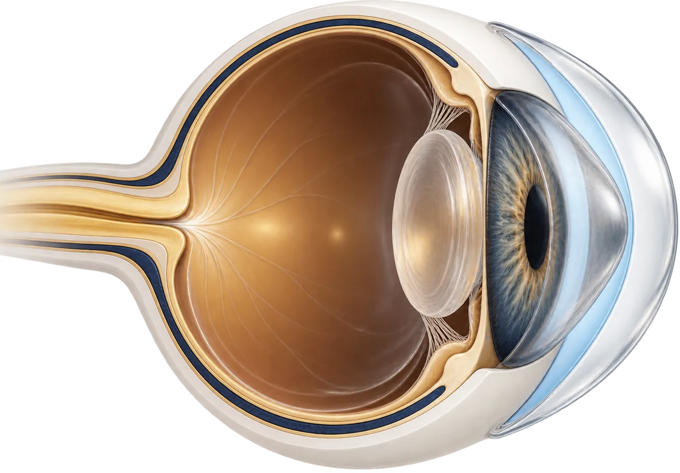
The cornea is the clear front window of the eye. It has to be both clear and smoothly curved to focus light. A scar can interfere with either, and often with both:
- It can warp the surface. As a wound or ulcer heals, the cornea can end up uneven, flattened in one area or pulled in another. This creates irregular astigmatism, which scatters light and causes blur, ghosting, and distortion that glasses can’t fully correct.
- It can cloud the tissue. Scar tissue isn’t as clear as healthy cornea. Where it sits in front of the pupil it blocks and scatters light, which causes haze and glare.
Which of these matters more depends on where the scar is, how deep it goes, and how it healed. That distinction decides how much a scleral lens can help.
Common causes of corneal scars include:
- Injury. Cuts, penetrating injuries, and the stitches used to repair them. Eye injury and corneal ulcers together are estimated to cause 1.5 to 2.0 million new cases of blindness in one eye every year worldwide, and the authors noted these causes are often underreported.[1]
- Bacterial infection. Bacterial corneal ulcers, including those linked to contact lens wear, can leave a scar after the infection clears.[6]
- Herpes simplex virus. Herpes keratitis can cloud the cornea and is believed to be the leading infectious cause of blindness in the developed world. One estimate puts its global incidence at about 1.5 million cases a year, including 40,000 new cases of severe vision loss in one eye.[2] It can come back: in a large trial, stromal keratitis recurred within 12 months in 28% of people on placebo and 14% of those taking preventive acyclovir.[4]
- Acanthamoeba. This amoeba causes a rare, hard-to-treat corneal infection. In one study, Acanthamoeba ulcers took a median of 113 days for the surface to heal, compared with 25 days for bacterial ulcers.[3]
- Surgery. Corneal surgery, including refractive surgery and transplants, can leave an irregular surface. In a large series of scleral lens fittings at one US center, 70 of 262 eyes with irregular corneas had irregularity left after surgery.[8]
How a scleral lens works on a scarred cornea
A scleral lens is a large rigid gas-permeable lens. It rests on the sclera (the white of the eye) and arches over the entire cornea without touching it. Before insertion it is filled with sterile saline, so a layer of fluid sits between the lens and the cornea.
The fluid fills in the dips and ridges of an uneven scarred surface. Light then passes through the smooth front of the lens and an even layer of fluid, which largely cancels out the surface irregularity. Because the lens rests on the white of the eye, it doesn’t have to balance on a lumpy or lopsided cornea the way a small rigid lens does.
What the fluid layer can’t do is make cloudy tissue clear. It works at the surface. If the scar has clouded tissue deeper in the cornea, in the line of sight, that haze stays exactly where it is with the lens in. The only way to remove deep scar tissue is surgery that removes or replaces it, usually a corneal transplant.
In practice, many scars cause both problems. A scleral lens can still help a great deal by removing the surface part of the blur. In one case report, a woman was left with a scar, an irregular surface, and some cloudiness in the pupil area after a bacterial infection. A scleral lens masked the surface problems and her vision improved substantially despite the cloudiness.[6]
What the research shows
Vision improves in eyes with scars. In a Swiss series of six eyes with scars in the line of sight, average vision was 20/80 with glasses and 20/25 with mini-scleral lenses. The average gain was five lines on the eye chart.[5] The authors noted that several of these scars also made the corneal surface markedly irregular.
The same holds after serious eye injuries. In a Turkish study of 11 people whose eyes had been repaired after open globe injuries, and who couldn’t tolerate smaller rigid lenses, vision improved significantly with scleral lenses: from a median of 0.50 to 0.10 on the logMAR scale, where lower is better.[7]
Larger series include scars among other irregular corneas. A US academic center reviewed 825 eyes fitted with one type of scleral lens. Of the 262 eyes with irregular corneas, 57 had corneal scars. Vision in the irregular cornea group improved by an average of 0.46 on the logMAR scale.[8]
These studies are small, mostly single-clinic, and without comparison groups. They show that scleral lenses often help, not how much they will help a particular scar.
Other options, compared
For scars close to the surface, a laser procedure called phototherapeutic keratectomy (PTK) can remove some scar tissue and smooth the surface. A review described it as highly effective for superficial corneal scars and noted it can improve vision even when the whole opacity can’t be removed.[11]
For deep or dense scars, a corneal transplant replaces the damaged tissue. Infection is a leading reason for transplants in some parts of the world. A review of 180,865 transplants found keratitis was the most common reason in Asia, at 32.3% of cases.[12] A transplant doesn’t always end the need for a specialty lens. Scleral lenses are also fitted after transplants.[10]
| Option | What it does | Typically suited to |
|---|---|---|
| Glasses | Correct regular blur only | Scars with little surface distortion |
| Soft or specialty soft lenses | Mask mild irregularity | Mild surface distortion |
| Corneal rigid gas-permeable lenses | Give a smooth optical surface on the cornea | Moderate irregularity where the lens centers and is comfortable |
| Scleral lenses | Vault the whole cornea over a fluid layer | Marked surface irregularity, or failure of the options above |
| Phototherapeutic keratectomy (PTK) | Laser removal and smoothing of surface tissue | Scars close to the surface |
| Corneal transplant (partial or full thickness) | Replaces scarred tissue | Deep or dense scars in the line of sight |
Questions to ask a scleral lens fitter
- How much of my blur do you think comes from the surface shape, and how much from the scar itself?
- Can we try a diagnostic lens first to see how much my vision improves?
- How many patients with corneal scars do you fit?
- How many visits does a fit usually take, and what do the fees cover?
- Do you bill my insurance directly, or give me a superbill to submit myself?
- Will you coordinate with my cornea specialist, especially if I’ve had herpes or Acanthamoeba?
- What signs mean I should remove the lens and call you the same day?
Common questions
Can a scleral lens make a corneal scar go away?
No. A scleral lens doesn't change the scar. It smooths out the uneven surface a scar can leave, which often improves vision a great deal. If the scar also clouds the tissue deep in the cornea, that cloudiness is still there with the lens in.
How do I know whether my blur is from the surface or from the scar itself?
Your eye doctor can often tell from an exam and corneal imaging. A trial with a rigid or scleral lens is also a practical test: if vision sharpens a lot with the lens, much of the blur was coming from the surface shape.
Can I wear scleral lenses after herpes keratitis?
Many people with herpes scars wear specialty lenses, but herpes can come back. Your cornea specialist should be involved in the decision and may recommend antiviral medicine to lower the chance of a recurrence.
Is a corneal transplant better than a scleral lens for a scar?
They solve different problems. A scleral lens corrects the surface shape without surgery. A transplant replaces the scarred tissue and is the only way to remove deep cloudiness, but it carries surgical risks and a long recovery. Many people try lenses first.
Can laser treatment remove a scar?
Some scars near the surface can be treated with a laser procedure called phototherapeutic keratectomy. It isn't suitable for deep scars. Your cornea specialist can tell you whether a scar is shallow enough.
How long should I wait after an injury or infection before getting fitted?
That is your eye doctor's decision. A new scar can keep changing for months as it settles, and lenses ordered too early may need replacing. Wearers often say waiting until the cornea is stable saved them a refit.
My vision is much better but still not perfect. Is this as good as it gets?
It may be, or there may be more to gain. Ask your fitter whether the remaining blur comes from haze deep in the scar, which a lens can't clear, or from leftover distortion, which fit changes or custom optics can sometimes reduce. If you're unsure, a second opinion from an experienced scleral fitter is reasonable.
Why does my lens fog so quickly over a scarred cornea?
A scarred or irregular cornea can be more prone to debris collecting in the saline under the lens. Changes to the fit or the fill sometimes help, and so does treating any surface inflammation. Tell your fitter how soon the fog starts.
Can the lens catch on my scar when I take it out?
It can happen if the edge drags across the cornea. Hold your lids wide so the lens comes away cleanly, and ask your fitter to watch your removal technique. If your eye hurts or stays red after taking the lens out, call your fitter.
Why is my vision worse in dim light?
In dim light your pupil widens, so light passes through more of the cornea, including scarred areas, which can add glare and blur. Many people with scars notice this most when driving at night. Tell your fitter, and ask whether anything in the lens design could help.