
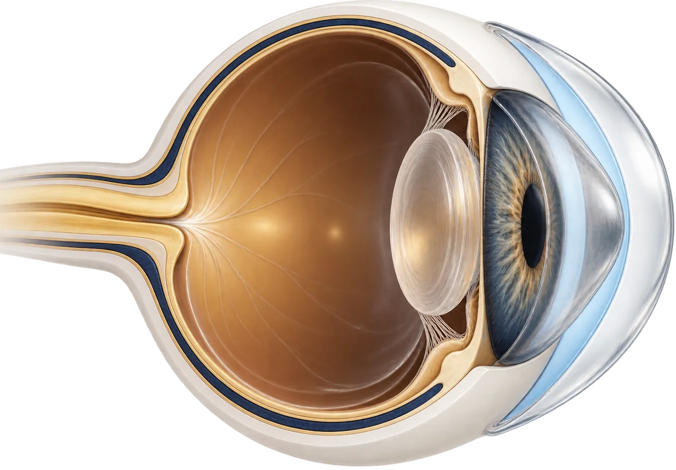
How neurotrophic keratitis changes the eye
The cornea (the clear front window of the eye) is supplied by branches of the trigeminal nerve. Those nerves do more than sense touch. They help the cornea’s surface layer stay healthy and repair itself. Neurotrophic keratitis (also called neurotrophic keratopathy) is what happens when that nerve supply is damaged and the cornea loses sensation. The surface breaks down on its own and heals poorly.[1]
Many conditions can damage the nerve. Herpes infections of the eye, diabetes, and eye or brain surgery are among the causes that share this mechanism.[1] On average, neurotrophic keratitis develops in about 6% of herpes simplex eye infections and 12.8% of shingles infections of the cornea.[1]
It is rare. Neurotrophic keratitis is classified as an orphan disease, with an estimated prevalence of fewer than 5 in 10,000 people.[1]
Doctors usually describe it in three stages:[1]
- Stage 1: changes in the surface layer of the cornea.
- Stage 2: a persistent epithelial defect, a patch of missing surface cells that won’t close.
- Stage 3: a corneal ulcer, which can lead to thinning, melting, and perforation.
One feature makes it treacherous. Because the cornea is numb, people with neurotrophic keratitis rarely complain of symptoms, even when the damage looks serious under the microscope.[1] Vision can drop as the surface breaks down or scars.
How a scleral lens works on a numb cornea
A scleral lens is a large rigid gas-permeable lens that rests on the sclera (the white of the eye) and arches over the entire cornea without touching it. It is filled with sterile saline before it goes in. That fluid stays against the cornea for as long as the lens is worn.
For a cornea that can’t protect or heal itself, this does three things:
- It keeps the surface constantly wet. The saline reservoir bathes the cornea all day, so it doesn’t depend on a tear film or blinking to stay moist.
- It shields the surface from the eyelids. Each blink drags the lid across the cornea. Over a fragile or open defect that friction can strip away new cells. The lens takes the rubbing instead.
- It can sharpen vision. The smooth lens surface and fluid layer mask an uneven or scarred cornea.
Researchers who treated stubborn defects this way concluded that healing seemed to come from the combination of oxygen, moisture, and protection that the lens provides.[3] PROSE (prosthetic replacement of the ocular surface ecosystem) is one scleral lens treatment model used in much of this research.
What the research shows
There are no randomized trials of scleral lenses for neurotrophic keratitis. A 2021 review found none comparing contact lenses with other treatments. The evidence comes from case reports and small case series, which suggest scleral lenses can be safe and effective at any stage of the disease.[2]
Defects that failed other treatments have healed. In a New York series, eight eyes with severe ocular surface disease and defects that hadn’t healed with standard treatment wore a PROSE lens around the clock under a strict protocol. The lens was removed briefly each day for cleaning, and an antibiotic drop was added to the reservoir. All eight defects healed and none of the eyes developed an infection.[5] In a Houston series of nine patients whose causes included neurotrophic keratopathy, all defects healed with PROSE after failing to heal with bandage soft lenses. Vision improved in eight of the nine.[4]
The benefit often lasts only as long as the lens is worn. In that Houston series, eight of nine patients had their defects come back when PROSE was stopped. All healed again once they resumed it.[4] In the New York series, four eyes had recurrences, and all responded when continuous wear restarted.[5]
Commercially available scleral lenses are used too. At the Mayo Clinic, neurotrophic keratopathy was one of the three most common reasons for fitting scleral lenses for ocular surface disease. Of 115 patients who completed fitting, all but 2 met their treatment goals, such as better comfort, surface protection, or healing.[6]
Long-term use is possible. A Belgian report followed three patients with a numb cornea and eyelids that didn’t close fully after brain tumor surgery. They wore scleral lenses for 3, 17, and 18 years as an alternative to having their eyelids partly sewn shut.[7]
Other options, compared
Scleral lenses are one of several treatments, and they are often combined with others.
Cenegermin (Oxervate) is an eye drop containing a lab-made version of human nerve growth factor. The FDA approved it in 2018 for neurotrophic keratitis. It is used six times a day for eight weeks.[8] In a US trial of 48 patients across 11 sites, 65.2% of patients on cenegermin had complete corneal healing at eight weeks, compared with 16.7% on placebo drops.[9][8] Among patients who healed, recurrences occurred in about 14% to 20% across the two trials.[8] The label says contact lenses should come out before each dose and can go back in 15 minutes later.[8]
Corneal neurotization is surgery that reroutes a healthy nerve, or a nerve graft, to the numb cornea. A 2025 review of 17 studies covering 232 eyes found it improved corneal sensation and vision.[10]
Amniotic membrane transplantation, tarsorrhaphy, and conjunctival flaps are surgical ways to protect the surface. They can preserve the eye but don’t restore sensation or improve vision.[1]
| Option | What it does | Typically suited to |
|---|---|---|
| Preservative-free lubricants | Support the tear film in place of preserved drops | Every stage, as a baseline |
| Bandage soft lens | Covers a defect short term | Early defects, with close monitoring |
| Scleral lens | Holds saline over the cornea and shields it from the lids | Persistent defects, or long-term protection with better vision |
| Cenegermin (Oxervate) drops | Nerve growth factor that promotes healing | Persistent defects or ulcers |
| Amniotic membrane | Tissue graft placed over the cornea to aid healing | Defects not responding to other treatment |
| Tarsorrhaphy | Partly closes the eyelids | Defects, especially with poor lid closure |
| Corneal neurotization | Restores a nerve supply to the cornea | Selected patients with lasting loss of sensation |
Questions to ask a scleral lens fitter
- How many patients with neurotrophic keratitis or persistent epithelial defects do you treat?
- Will you work with my cornea specialist on the treatment plan?
- Should the lens be worn overnight while my defect heals, or only during the day?
- Will I use an antibiotic while a defect is open, and how often will you check for infection?
- How do I fit my cenegermin drops or other medicines around the lens?
- If I can’t feel my eye, what signs should make me remove the lens and call right away?
Common questions
Can a scleral lens bring back feeling in my cornea?
No. A scleral lens protects the surface and keeps it wet, but it doesn't repair the nerve. Treatments aimed at the nerve itself include cenegermin eye drops and corneal neurotization surgery.
Why do I need to be checked so often?
Contact lens wear may raise the risk of infection in a cornea with an open defect, and a numb eye may not hurt the way an infected eye normally would. Frequent checks catch infection early. Your doctor may also add an antibiotic while a defect is open.
Can I use Oxervate drops while wearing a scleral lens?
The drug's label says to take contact lenses out before each dose and wait 15 minutes before putting them back in. Ask your doctor how to schedule the drops around your lens.
Will I have to wear the lens forever?
Possibly. In small studies, epithelial defects often came back when lens wear stopped and healed again when it resumed. Some people use the lens long term to keep the surface stable.
Is a scleral lens better than having my eyelids partly sewn shut?
They suit different people. Tarsorrhaphy is simple and effective but narrows your vision and changes how the eye looks. A scleral lens keeps the eye open and can improve sight, but takes practice to handle. Your cornea specialist can help you weigh them.
Can a scleral lens help my cornea heal?
It can. In small studies, surface defects that hadn't healed with other treatments often closed with a scleral lens, because the lens keeps the cornea wet and shields it from the eyelids. Your doctor decides how the lens is used, sometimes with an antibiotic, and will check the eye closely while it heals.
My eye is numb. What warning signs should I watch for?
Don't wait for pain, because a numb eye may not hurt even when something is seriously wrong. Watch for redness, discharge, blurrier vision, light sensitivity, or a white spot on the eye. If you notice any of these, remove the lens and call your eye doctor today.
Can I use serum tears and a scleral lens together?
Many people with corneal nerve damage use both, and some wearers say the lens helped more than drops alone. Your doctor will tell you when to use the drops around your lens wear. Adding drops into the lens fill is off-label, so only do that if your doctor tells you to.
Is it harder to put the lens in when my eye can't feel it?
It can be, because you may not feel a bubble or a lens that has landed off-center. Check each lens with a mirror and good light before and after it goes in, and look for bubbles. Ask your fitter to teach you how to check your lens by sight.