
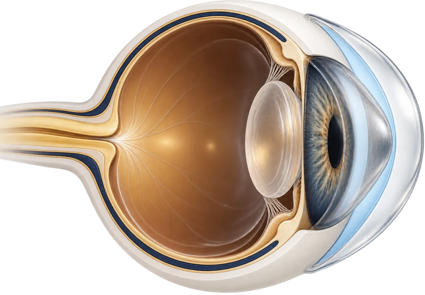
How post-LASIK ectasia changes your vision
Laser vision surgery reshapes the cornea (the clear front window of the eye) by removing tissue. In LASIK the surgeon first cuts a thin flap, in PRK the surface layer is removed, and in SMILE a small disc of tissue is taken out through a small incision. In most eyes the thinner cornea stays stable for life.
In a small number, it doesn’t. The cornea weakens, thins, and bulges forward, much as it does in keratoconus. This is called ectasia. It typically brings increasing nearsightedness and astigmatism.[7] The surface also becomes irregular, so light is scattered instead of focused. People describe blur, ghosting, and a prescription that keeps changing, sometimes years after surgery gave them clear vision without glasses.
Glasses correct regular blur. They can’t correct irregular distortion, which is why vision in glasses often stays poor once ectasia sets in.
How often it happens depends on who’s counting. A 2021 review of published reports estimated ectasia in 90 per 100,000 LASIK eyes, 20 per 100,000 PRK eyes, and 11 per 100,000 SMILE eyes, counting only eyes with no known risk factors beforehand. By that count the rate after LASIK was 4.5 times the rate after PRK.[1] One hospital that tracked 30,167 LASIK eyes for up to eight years found ectasia in 10 of them, or 0.033%.[2] A 2001 report had put the figure at 0.66%, and screening has improved a great deal since then.[3] For SMILE, one estimate is 0.011%, though the procedure is newer and there is less data behind it.[4] Estimates vary with the procedure, the era, and how cases were found.
It doesn’t always show up right away. In one US case series, ectasia appeared an average of 16.3 months after LASIK, with a range of 1 to 45 months.[5] Another study noted that some cases may appear beyond eight years.[2]
Most cases have a risk factor in hindsight. In that same US series, 88% of eyes that developed ectasia had met criteria for very early (forme fruste) keratoconus before surgery. High nearsightedness and a thin layer of untouched cornea left under the flap were the other significant risk factors.[5] A risk-scoring system built from these factors correctly flagged 46 of 50 ectasia eyes (92%) as high risk in a later study.[6] Even so, some cases occur with no identifiable risk factor. Eye rubbing and ocular allergy may play a part.[3][2]
How a scleral lens works on an ectatic cornea
A scleral lens is a large rigid gas-permeable lens. Instead of sitting on the cornea, it rests on the sclera (the white of the eye) and arches over the entire cornea without touching it. Before insertion the lens is filled with sterile saline, so a layer of fluid sits between the lens and the cornea.
That arrangement does two things:
- It replaces the eye’s irregular front surface. Light passes through the smooth front of the lens and the even layer of fluid before it reaches the cornea. The fluid fills in the cornea’s irregularities, which largely cancels them out optically.
- It keeps the lens off the weakened area. Smaller rigid lenses sit directly on the cornea and can be hard to keep centered on an irregular surface. A scleral lens lands on the white of the eye instead, so the shape of the cornea matters much less for fit.
Rigid lenses have long been the usual way to restore vision in ectasia. In the US case series above, one eye out of ten needed a corneal transplant. All the others were corrected with rigid gas-permeable contact lenses.[5]
What the research shows
Studies of post-LASIK ectasia specifically are small. Much of the evidence comes from case series and from studies that mix several corneal conditions together. A 2015 review found high reported success with scleral lenses after refractive surgery, but noted that outcomes for refractive surgery patients were not reported separately.[9]
Scleral lenses sharpen vision and reduce distortion. A study in India fitted one scleral lens design to 46 eyes that couldn’t see well enough in glasses or soft lenses. 19 of them had post-LASIK ectasia. With the lens in, median corrected vision improved from 0.30 to 0.0 logMAR (a lower number is sharper), and median astigmatism fell from 3.75 to 0.50 diopters. Higher-order distortion also dropped significantly.[8]
Most people in mixed studies keep wearing them. In a prospective study of 40 eyes with a range of corneal problems, including eyes after refractive surgery, people wore their mini-scleral lenses an average of 12.1 hours a day, and 12.5% stopped wearing them. Follow-up was only three months.[10]
The cornea can still change under the lens. A Spanish study followed 20 eyes with post-LASIK ectasia for a year of scleral lens wear. Corneal thickness and curvature changed measurably, even though vision with the lens stayed the same. The authors recommended regular corneal mapping for people wearing these lenses.[11]
The long-term outlook with lenses can be good. In the US case series, every patient eventually reached corrected vision of 20/30 or better, and only 10% of eyes lost more than one line of best-corrected vision.[5]
Cross-linking and other options
Corneal cross-linking strengthens the cornea to slow or stop ectasia progressing. The FDA approved the first cross-linking system in the US in 2016, for both progressive keratoconus and corneal ectasia following refractive surgery.[13]
In the US trial behind that approval, 179 people with ectasia after refractive surgery were randomly assigned to cross-linking or a sham treatment. After one year, peak corneal steepness had fallen by 0.7 diopters in the cross-linked eyes and kept rising in the untreated eyes, a 1.3-diopter difference. 32% of treated eyes gained 10 or more letters of corrected vision.[12] Corneal haze was the most frequently reported side effect.[12] In 6% of ectasia patients in the approval studies, haze was still present at 12 months.[13] Cross-linking has also been used for ectasia after SMILE.[4]
Cross-linking and scleral lenses solve different problems. Cross-linking aims to keep the cornea from getting worse. A lens is usually still needed for clear vision afterward.
| Option | What it does | Typically suited to |
|---|---|---|
| Glasses | Correct regular blur only | Mild ectasia with little irregularity |
| Soft or specialty soft lenses | Mask mild irregularity | Mild cases |
| Corneal rigid gas-permeable lenses | Give a smooth optical surface on the cornea | Mild to moderate cases where the lens centers and is comfortable |
| Hybrid lenses | Rigid center with a soft skirt | People who can’t tolerate a corneal rigid lens |
| Scleral lenses | Vault the whole cornea over a fluid layer | Moderate to severe cases, or failure of the options above |
| Corneal cross-linking | Strengthens the cornea to slow or stop progression | Progressive ectasia; doesn’t replace vision correction |
| Corneal transplant | Replaces corneal tissue | Scarring or intolerance of every lens option |
Questions to ask a scleral lens fitter
- How many patients with ectasia after refractive surgery do you fit?
- How do you measure my eye: trial lenses, corneal mapping, or a 3D scan of the eye surface?
- How often will you map my cornea to check for changes while I wear the lens?
- How many visits does a fit usually take, and what do the fees cover?
- Do you bill my insurance directly, or give me a superbill to submit myself?
- Who do I call if I can’t get a lens out?
- Should I be evaluated for cross-linking before or alongside the fitting?
Common questions
I had LASIK years ago. Can ectasia still happen?
It can appear months or years after surgery. In one US case series the average was about 16 months after surgery, with a range of 1 to 45 months, and some cases may appear later still. If your vision has been getting worse since surgery, tell your eye doctor you had refractive surgery.
Can scleral lenses stop ectasia from getting worse?
No. A scleral lens changes how light enters the eye, not the strength of the cornea. Corneal cross-linking is the treatment designed to slow or stop progression. Many people have cross-linking and then wear a scleral lens for vision.
Can I have more laser surgery to fix it?
Ectasia is a weakening of the cornea, so any further surgery needs careful assessment. Your surgeon will talk you through which options are safe for your eye. Cross-linking and specialty lenses are the options most often discussed.
Is post-LASIK ectasia the same as keratoconus?
They look and behave alike, and they are managed in similar ways. Many people who develop ectasia after surgery had signs of very early keratoconus beforehand that weren't recognized at the time.
Can ectasia happen after PRK or SMILE too?
Yes, though reported rates are lower than after LASIK. Ectasia has been reported after all three procedures. Estimates vary, and newer procedures have less long-term data behind them.
My records say post-LASIK ectasia. Should I be worried?
It's the medical name for the corneal weakening and bulging that can follow laser surgery, and it explains why your vision changed. It's a condition that can be managed. Ask a cornea specialist whether it's progressing and whether cross-linking makes sense for you, and keep your follow-up visits.
Should I try glasses or soft lenses before scleral lenses?
If glasses or soft lenses give you vision you're happy with, they are simpler, and many fitters try the simplest workable option first. Once ectasia makes the cornea irregular, glasses often can't give clear vision. That is usually when a rigid or scleral lens is considered.
Will scleral lenses get rid of halos and starbursts at night?
For many people they reduce them a lot, but not always completely. Some distortion can remain because of the cornea's shape or a lens that sits slightly off-center. Tell your fitter what you see at night, and ask whether adjustments or custom optics designed to correct leftover distortion are an option for you.
Can a scleral lens also help with dry eye after LASIK?
Often, yes. The saline under the lens keeps the cornea bathed while you wear it, and many people who had laser surgery wear scleral lenses for both clearer vision and comfort. Dry eye usually needs its own treatment too, such as care for the eyelids and oil glands, so keep your dry eye doctor involved.
My first scleral lenses didn't help my vision much. Should I give up?
Not necessarily. Fitting an irregular cornea is complex, and it often takes several adjustments, a different design, or added optical correction to get the best result. Tell your fitter exactly what is wrong, and consider a second opinion from a fitter who sees many post-surgery eyes.