
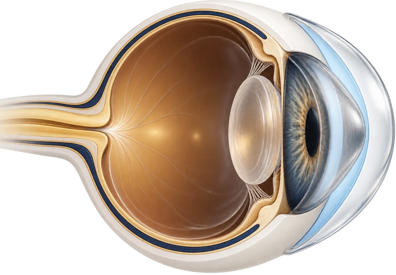
How pellucid marginal degeneration changes your vision
Pellucid marginal degeneration (PMD) is a rare condition in which a crescent-shaped band of the cornea, usually near its lower edge, becomes thin.[2] The cornea just above that band bulges forward. The result is a lot of irregular astigmatism, which blurs and distorts vision.
The astigmatism in PMD is often “against the rule.” That means the cornea is steeper across its horizontal meridian than its vertical one, the opposite of the usual pattern. Reviews describe increasing against-the-rule irregular astigmatism leading to a slow drop in vision as a typical sign.[1] In one study of rigid lens wearers with PMD, 63.3% had against-the-rule astigmatism, and the average astigmatism was more than 8 diopters.[5]
Corneal maps of PMD often show a distinctive “crab-claw” pattern.[2] Early PMD can go unnoticed without corneal mapping, and later PMD is sometimes mistaken for keratoconus.[1]
How common it is isn’t known. A 2011 review stated plainly that the prevalence and cause of PMD remain unknown.[1] We found no population-based prevalence study. The largest clinical series comes from an Indian hospital network, which diagnosed PMD in 559 of 2,470,793 new patients (0.02%) over about eight years.[4] That is a count of people who came to eye hospitals, not a survey of the general population, so it can’t tell you how common PMD is in any given country.
In that same series, 70.13% of patients were male, 77.1% had it in both eyes, and the average age was about 38.[4] A review describes it as most common in men, usually appearing between the second and fifth decades of life, and found in all ethnic groups.[1]
In rare cases, the thin area can swell suddenly (called hydrops) or even perforate, causing sudden pain and loss of vision.[1] In the Indian series, hydrops was seen in 1.01% of affected eyes.[4]
How a scleral lens works for PMD
A scleral lens is a large rigid gas-permeable lens. It rests on the sclera (the white of the eye) and arches over the entire cornea without touching it. Before insertion it is filled with sterile saline, so a layer of fluid sits between the lens and the cornea.
That design matters for PMD in two ways:
- It replaces the irregular front surface. Light passes through the smooth front of the lens and the fluid layer before reaching the cornea. The fluid fills in the cornea’s uneven shape, which largely cancels out the irregular astigmatism optically.
- It doesn’t depend on the cornea to hold it in place. A small rigid corneal lens sits on the cornea itself. In PMD the steepest area is low and close to the edge, so a corneal lens can ride low, rock, or pop out. A scleral lens lands on the white of the eye, beyond the thin band, so the cornea’s shape matters much less for fit.
Many people with PMD do well in rigid corneal lenses, especially larger ones. In one study, 24 of 27 eyes were successfully fitted with rigid gas-permeable lenses, and the authors found larger-diameter lenses were better tolerated.[5] Scleral lenses are often used when those don’t work. In one series, the reasons for moving to a scleral lens included lenses popping out and failure of other rigid lens approaches.[7]
What the research shows
The studies on scleral lenses for PMD are small case series: the ones below range from 9 to 24 eyes.[9][8] They consistently show better vision, but they can’t tell you how well any one person will do.
Vision improves in most eyes fitted. In a Turkish study of 24 eyes with PMD, vision with scleral lenses was on average 3.3 lines better than with glasses.[8] A French series of 9 eyes found vision improved from 0.51 to 0.04 on the logMAR scale, where lower is better.[9] An Indian series of 20 eyes found a similar, significant improvement.[7]
Astigmatism and distortion drop. A study of 46 eyes with PMD, keratoconus, or ectasia after LASIK measured distortion before and during scleral lens wear. Across all groups, the median astigmatism in the prescription fell from 3.75 to 0.50 diopters, and higher-order distortion fell significantly.[10]
Most PMD is managed without surgery. In the Indian hospital series, 87.97% of eyes were managed with glasses or contact lenses.[4] A British study found 80% of non-surgically managed eyes saw 6/12 (20/40) or better after about 32 months, and the authors concluded that scleral-fitted gas-permeable lenses can correct PMD well over the long term.[6] A 2025 review concluded that most PMD patients can be managed with rigid contact lenses.[3]
Other options, compared
Cross-linking, a procedure that strengthens corneal tissue, has been used to try to stabilize PMD. A 2019 review found it may halt progression, but noted the data are limited and mostly short-term and retrospective.[11] In the Indian series, it was the most common surgery performed, in 5.25% of eyes.[4]
Surgery for PMD has mixed results. In the British study, the average astigmatism after wedge excision surgery was still 8.90 diopters, and the authors described the long-term reduction in astigmatism from surgery as poor.[6] Reviews reserve transplants for severe PMD and note risks including graft rejection and astigmatism after surgery.[2] Lamellar (partial-thickness) transplants appear to give results similar to full-thickness transplants with fewer complications.[3]
| Option | What it does | Typically suited to |
|---|---|---|
| Glasses | Correct regular astigmatism only | Early PMD with little irregularity |
| Soft or toric soft lenses | Mask mild irregularity | Mild cases |
| Corneal rigid gas-permeable lenses | Give a smooth optical surface on the cornea; larger designs center better | Mild to moderate cases where the lens stays in place |
| Hybrid lenses | Rigid center with a soft skirt | People who can’t tolerate a corneal rigid lens |
| Scleral lenses | Vault the whole cornea over a fluid layer | Moderate to advanced cases, or when smaller lenses decenter or fall out |
| Corneal cross-linking | Strengthens corneal tissue to try to stop progression | Progressive PMD; evidence still limited; doesn’t replace vision correction |
| Corneal transplant or wedge excision | Replaces or removes thinned tissue | Advanced cases where lenses no longer work |
Questions to ask a scleral lens fitter
- How many patients with PMD do you fit, and how does it change your approach compared with keratoconus?
- How do you measure my eye: trial lenses, corneal mapping, or a 3D scan of the eye surface?
- How will you check that the lens clears the thin area near the bottom of my cornea?
- How many visits does a fit usually take, and what do the fees cover?
- Do you bill my insurance directly, or give me a superbill to submit myself?
- What signs mean I should remove the lens and call you the same day?
- Should I be evaluated for cross-linking?
Common questions
Is pellucid marginal degeneration the same as keratoconus?
No, though they are related. Both thin and reshape the cornea. In PMD the thinning is a narrow band near the lower edge of the cornea, while in keratoconus the cone is usually more central. Later-stage PMD is sometimes mistaken for keratoconus.
How common is pellucid marginal degeneration?
Nobody knows for certain. There are no good population studies, so reliable prevalence figures don't exist. It is considered rare. One large Indian hospital network diagnosed it in 559 of nearly 2.5 million new patients over eight years.
Why do small rigid lenses keep popping out?
The steepest part of a PMD cornea sits low, near the edge. A small lens that rests on the cornea can ride low, rock, or fall out. A scleral lens rests on the white of the eye instead, so the cornea's shape matters less for how it stays in place.
Can scleral lenses stop PMD from getting worse?
No. A scleral lens changes how light enters the eye, not the course of the disease. Cross-linking has been used to try to stabilize PMD, but the evidence for it is still limited. Ask your eye doctor whether it applies to you.
Will I need a corneal transplant?
Most people with PMD are managed with glasses or contact lenses. Transplants are usually reserved for severe cases and carry risks, including graft rejection and astigmatism after surgery. Your eye doctor can tell you where your eyes stand.