
How SJS and TEN affect the eye and vision
Stevens-Johnson syndrome (SJS) and toxic epidermal necrolysis (TEN) are severe reactions, most often to a medicine, that cause the skin and mucous membranes to blister and peel. Doctors tell them apart by how much skin detaches. In the standard classification, SJS involves detachment of less than 10% of the body surface, overlap SJS/TEN 10% to 30%, and TEN usually more than 30%.[1] Survivors can have lasting problems in the skin, mouth, lungs, gut, and eyes, and many also carry psychological effects from the illness.[2]
The eyes are involved often. In a French referral center, 74% of 159 patients with SJS or TEN had eye involvement during the acute illness. Among 49 patients contacted at least 15 months later, 63% had late eye complications, and dry eye was the most common. Some people developed dry eye even without obvious eye problems during the acute phase.[3]
The chronic eye problems come from scarring of the eye surface and eyelids:
- Lid margin disease. The lid margins scar and thicken, the oil glands stop working, and lashes can turn inward and rub the eye. In a study of patients treated with amniotic membrane during the acute illness, oil-gland disease and dry eye were still the most common long-term problems, affecting 78% and 58% of eyes.[4]
- Dryness. Damaged tear glands and oil glands leave the surface without a stable tear film.
- Scarring of the conjunctiva. Bands of scar tissue can tie the lid to the eyeball and shorten the pocket behind the lid.
- Corneal damage. Blood vessels and opaque tissue can grow over the cornea (the clear front window of the eye), and its surface can become skin-like and keratinized. This is what threatens vision most.
- Light sensitivity and discomfort. A dry, inflamed, irregular surface makes light glare painful and vision blurry.
The damage can keep getting worse long after the acute illness. In a study of 105 eyes followed for more than five years, surface scarring worsened in 33.3%. Eyes with more severe scarring of the lid margin and upper lid were more likely to get worse.[5]
How a scleral lens works after SJS or TEN
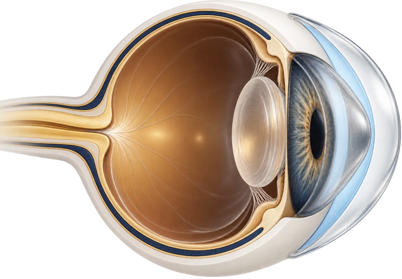
A scleral lens is a large rigid gas-permeable lens. It rests on the sclera (the white of the eye) and arches over the whole cornea without touching it. Before insertion the lens is filled with sterile, preservative-free saline, so a pool of fluid sits against the cornea for as long as the lens is in.
That arrangement helps an SJS eye in three ways:
- It keeps the cornea bathed. When the eye can no longer make a healthy tear film, the saline reservoir keeps the surface wet all day.
- It shields the cornea from the lids. Scarred lid margins and misdirected lashes rub the cornea with every blink. With a scleral lens in place they rub the lens instead.
- It smooths the optics. The fluid layer fills in an irregular corneal surface, so light is focused more evenly and vision often sharpens.
PROSE (prosthetic replacement of the ocular surface ecosystem) is a scleral-device treatment developed by the Boston Foundation for Sight. Several of the larger SJS studies used PROSE devices. Other studies used custom scleral lenses.
What the research shows
Vision improves in most people who can be fitted. A study of scleral device treatment followed 167 eyes of 86 patients with a history of SJS/TEN. Median vision improved from 20/60 at the first visit to 20/25 once the device was customized, with no decline at the end of follow-up. Median follow-up was 16 months. Scores on a standard visual-function questionnaire rose from a mean of 48 to 72 at six months.[6]
Quality of life improves too. In a study of 67 eyes of 39 patients, the mean visual-function score rose from 25.1 to 67.4 six months after scleral lens fitting. The mean Ocular Surface Disease Index (a symptom score, where higher is worse) fell from 76.9 to 37.1. Fitting failed in 3 patients, and no serious side effects from the lenses were seen.[7] A smaller Los Angeles study of 27 eyes also found significant gains in vision and symptom scores with PROSE.[8]
Children can benefit, though not all. A review of 49 children with SJS/TEN treated at one center found median vision improved from 20/80 at the first visit to 20/30 when the device was dispensed, and stayed there at the last visit. Mean follow-up was 5.45 years. Treatment failed in 15 children (30.6%).[9]
Scleral devices work best as part of a plan. A study of 568 eyes of 284 children with chronic SJS compared supportive care with definitive treatments. Definitive treatment, including PROSE, mucous membrane grafting of the lid margins, limbal stem cell transplants, and keratoprosthesis, improved vision and helped prevent corneal damage. In eyes where the lids were damaging the cornea, lid-margin grafting worked better than PROSE alone, and grafting followed by PROSE gave the best results.[10]
Other options, compared
Care after SJS/TEN usually combines several treatments. Some are for the acute illness, and others address the chronic damage. Your cornea specialist decides the mix.
| Option | What it does | Typically suited to |
|---|---|---|
| Amniotic membrane (acute phase) | Covers the eye surface during the acute illness to limit scarring | The first days to weeks of SJS/TEN, while in hospital |
| Artificial tears, ointments, and anti-inflammatory drops | Lubricate and calm the surface | Everyone with chronic eye involvement, as a base layer |
| Lash and lid procedures | Remove or redirect lashes that rub the eye | Misdirected lashes |
| Mucous membrane grafting of the lid margin | Replaces scarred, keratinized lid margin with tissue from inside the mouth | Lid margins that damage the cornea |
| Scleral lenses | Hold a saline reservoir over the cornea and shield it from the lids | Dry, irregular, or exposed corneas where there is room to fit a lens |
| Limbal stem cell procedures | Replace the stem cells that renew the corneal surface | Limbal stem cell deficiency with a conjunctivalized cornea |
| Keratoprosthesis (artificial cornea) | Replaces the central cornea with a clear optic | End-stage disease where other options have failed |
Early amniotic membrane treatment in the acute phase can reduce later vision loss. In the amniotic membrane study, vision was 20/40 or better in 87% of eyes at last follow-up.[4] Reviews stress that the earliest treatments are the most likely to prevent chronic complications, and that later treatments become more invasive and higher risk.[11] For long-term care, reviews list scleral lenses together with limbal stem cell transplantation, mucous membrane grafting, and keratoprosthesis.[12]
Questions to ask a scleral lens fitter
- How many patients with SJS or TEN do you fit each year?
- Is there enough room between my lids and my eye to fit a lens?
- Should my lid margins or lashes be treated before a fitting?
- Will you work with my cornea specialist on the overall plan?
- How many visits does a fit usually take, and what do the fees cover?
- Can a family member learn to insert and remove the lenses?
- Do you bill my insurance directly, or give me a superbill to submit myself?
Common questions
Can scleral lenses undo the damage from Stevens-Johnson syndrome?
No. Scarring of the lids, conjunctiva, and cornea stays. A scleral lens protects the surface, keeps it wet, and can smooth out an irregular cornea optically, which is why vision and comfort often improve. It doesn't restore healthy tissue.
Is it too late for scleral lenses if my SJS was years ago?
Not necessarily. Most of the published studies fitted people with chronic eye disease, often long after the acute illness. Whether a lens can be fitted depends on how much scarring there is between the lids and the eye, which a fitter checks in person.
Can children with SJS wear scleral lenses?
Yes, in some cases. In one series of 49 children, treatment worked in most, but failed in 30.6%. Children need a parent or caregiver who can handle the lenses and keep up with follow-up visits.
Will scleral lenses help with light sensitivity?
They may. Much of the glare and light sensitivity after SJS comes from a dry, damaged surface, and a lens that keeps the cornea wet can ease it. Sunglasses and tinted lenses are still commonly needed. Your eye doctor can tell you what is driving your symptoms.
Are scleral lenses covered by insurance after SJS or TEN?
Sometimes. Some medical plans cover scleral lenses when they are medically necessary for eye-surface disease. Coverage depends on your plan and on whether the practice is in network, so ask for the codes and a written estimate.
Can scleral lenses work if SJS left me with almost no tears or working oil glands?
Many SJS survivors with severe tear and oil gland loss wear scleral lenses, because the lens holds its own reservoir of saline over the cornea. Very dry eyes still make the lenses harder work, and many wearers keep using lubricating drops through the day. Your fitter can tell you how your eyes are likely to respond.
My lenses fog up after a few hours. What can help?
Fogging is common when the eye surface is damaged. Wearers report that fit changes, a different fill, and treating lid and surface inflammation can all reduce it, though what works varies. Adding drops to the fill is off-label, so make that choice with your fitter.
Is it normal to need several lenses before the fit is right?
Yes, especially when scarring has changed the shape of the lids and the white of the eye. Several remakes are common, and switching to a different design or fitter has helped some people after repeated failures. If you feel stuck, ask for a second opinion from a fitter experienced with SJS.
Do I need to go to a specialty center, or can a local fitter help?
Both routes work for different people. Specialty centers see many SJS eyes, while a local fitter may be easier to reach for the frequent visits a fit can need. What matters most is the fitter's experience with scarred eye surfaces, so ask how many SJS patients they see.
My vision has slowly gotten worse after years of wear. Is the lens to blame?
Not necessarily. SJS can keep changing the eye over time, for example through blood vessels growing into the cornea or loss of the cells that renew its surface, and other problems such as cataract can also blur vision. Ask your eye doctor and a cornea specialist to look for the cause, and tell your fitter what has changed.
Can I nap with my lenses in?
No. Take the lenses out before any sleep, including a short nap, unless your eye doctor has prescribed overnight wear for a specific reason. If you want protection for your eyes while you sleep, ask your eye doctor what to use instead.