
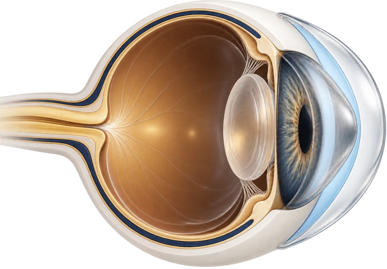
How keratoconus changes your vision
In keratoconus, the cornea (the clear front window of the eye) thins and bulges forward into a cone. A healthy cornea is a smooth, even curve. A keratoconic cornea is steeper in one area and irregular across its surface, so light entering the eye is scattered instead of focused. People describe ghosting, doubled images, halos around lights, and a prescription that keeps changing.
Glasses correct regular blur. They can’t correct the irregular distortion keratoconus causes, which is why vision in glasses often stays poor even with an up-to-date prescription.
How common it is depends on who’s counting. A Dutch study of insurance records covering 4.4 million people estimated about 1 in 375 people have keratoconus.[1] A worldwide review of 29 studies put the figure at 1.38 per 1,000.[2] An older study from Olmsted County, Minnesota, found 54.5 per 100,000.[3] Estimates vary widely with the population studied and how cases are found, so treat any single figure as a rough guide.
It usually appears young. In the Dutch registry the average age at diagnosis was 28.3, and 60.6% of those diagnosed were male.[1] Eye rubbing and a family history of keratoconus are the strongest known risk factors. Allergy, asthma, and eczema are also linked to it.[2] About half the patients in the CLEK study reported a history of allergic conditions.[4]
Keratoconus affects both eyes, even when one eye looks normal at first. A 2015 global consensus of cornea specialists concluded that true one-eyed keratoconus does not exist.[5][6] The two eyes are often affected unequally, though. In Olmsted County, 41% of patients appeared to have it in only one eye when they were diagnosed.[3]
How a scleral lens works on a keratoconic cornea
A scleral lens is a large rigid gas-permeable lens. Instead of sitting on the cornea, it rests on the sclera (the white of the eye) and arches over the entire cornea without touching it. Before insertion the lens is filled with sterile saline, so a layer of fluid sits between the lens and the cone.
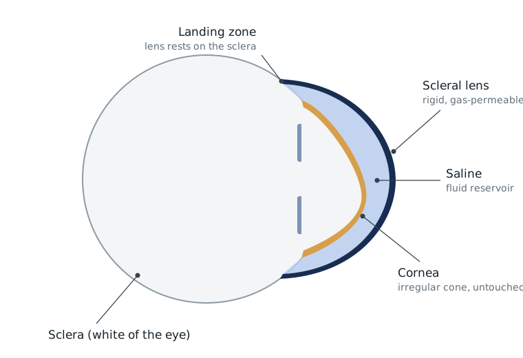
That arrangement does two things:
- It replaces the eye’s irregular front surface. Light passes through the smooth front of the lens and the even layer of fluid before it reaches the cornea. The fluid fills in the cornea’s irregularities, which largely cancels them out optically.
- It keeps the lens off the cone. Smaller rigid lenses sit on the cornea and can rub the steepest point. They can also be hard to keep centered on a very irregular cornea. A scleral lens lands on the white of the eye instead, so how steep the cone is matters much less for fit and comfort.
Rigid lenses have long been the standard vision correction for keratoconus. In the CLEK study of 1,209 patients, 65% wore rigid gas-permeable lenses.[4] Scleral lenses are the version that vaults the cornea entirely. Keratoconus and other corneal irregularity are their main use: in a 2020 survey of scleral lens fitters, a median of 84% of the scleral lenses fitted were for corneal irregularity.[13]
What the research shows
Scleral lenses can keep people with severe keratoconus from needing a transplant. A Belgian clinic followed 51 eyes with severe keratoconus that would otherwise have been referred for transplant surgery. After about two and a half years, 40 of those eyes were still wearing scleral lenses successfully. The authors reported that scleral lenses more than halved the need for transplants in their clinic.[11]
They improve day-to-day vision and quality of life. In a study of 89 eyes, mini-scleral lenses significantly improved both vision and patients’ own ratings of how their vision affected daily life.[12]
Transplants for keratoconus have been falling. In the long-running CLEK study, 12% of patients had a transplant in at least one eye over eight years.[8] Rates are higher at specialist hospital clinics that see more advanced cases: 21.6% of patients at one London hospital’s keratoconus clinic had been grafted by the end of that study.[9] In the US, keratoconus was the sixth most common reason for a corneal transplant in 2020. The Eye Bank Association of America attributes its steady decline to cross-linking and modern rigid and scleral lenses.[10]
Scleral lenses and cross-linking work together
Corneal cross-linking is a procedure that strengthens the cornea to stop keratoconus progressing. The FDA approved the first cross-linking system in the US in 2016, for progressive keratoconus.[14]
Cross-linking and scleral lenses solve different problems:
- Cross-linking aims to stop the cone getting worse. In the two trials behind the FDA approval, the steepest part of the cornea flattened on average by only 1.4 to 1.7 diopters at one year.[14] That is small next to the distortion many patients have.
- Scleral lenses provide the clear vision. For many people cross-linking stabilizes the eye, and a scleral lens is how they then see well.
Cross-linking works. In a randomized trial of 10- to 16-year-olds, keratoconus progressed in 7% of cross-linked eyes compared with 43% of eyes given standard care.[15] After the Netherlands introduced cross-linking, it performed about 25% fewer transplants for keratoconus over the next three years.[16]
Other options, compared
| Option | What it does | Typically suited to |
|---|---|---|
| Glasses | Correct regular blur only | Early keratoconus with little irregularity |
| Soft or specialty soft lenses | Mask mild irregularity | Mild cases |
| Corneal rigid gas-permeable lenses | Give a smooth optical surface on the cornea | Mild to moderate cases where the lens centers and is comfortable |
| Hybrid lenses | Rigid center with a soft skirt | People who can’t tolerate a corneal rigid lens |
| Scleral lenses | Vault the whole cornea over a fluid layer | Moderate to severe cases, or failure of the options above |
| Corneal cross-linking | Strengthens the cornea to stop progression | Progressive keratoconus; doesn’t replace vision correction |
| Corneal transplant | Replaces corneal tissue | Scarring or intolerance of every lens option |
Questions to ask a scleral lens fitter
- How many scleral lens patients with keratoconus do you see?
- How do you measure my eye: trial lenses, corneal mapping, or a 3D scan of the eye surface?
- How many visits does a fit usually take, and what do the fees cover?
- Do you bill my insurance directly, or give me a superbill to submit myself?
- Who do I call if I can’t get a lens out?
- Should I be evaluated for cross-linking before or alongside the fitting?
Common questions
Can scleral lenses stop keratoconus from getting worse?
No. A scleral lens changes how light enters the eye, not the course of the disease. Corneal cross-linking is the treatment designed to stop progression. Many people have cross-linking and then wear scleral lenses for vision.
Do scleral lenses press on the cone?
They are designed not to. The lens rests on the white of the eye and arches over the cornea with a layer of fluid in between. Your fitter checks that clearance at each visit.
Can I wear scleral lenses after cross-linking?
Yes, this is common. Your surgeon and fitter will tell you how long to wait after the procedure before a fitting.
Will I need a corneal transplant eventually?
Most people with keratoconus don't. In the long-running CLEK study, 12% had a transplant in at least one eye over 8 years. Transplant rates have fallen since cross-linking and modern specialty lenses became available.
Are scleral lenses covered by insurance for keratoconus?
Sometimes. Some medical and vision plans cover contact lenses when they are medically necessary, which can include keratoconus. Coverage depends on your plan and on whether the practice is in network.
Do I need scleral lenses if my keratoconus is mild and glasses still work?
Not necessarily. If glasses or soft lenses give you vision you are happy with, there may be no reason to switch yet. Scleral lenses become an option when glasses can no longer give you clear, comfortable vision. Your eye doctor can help you decide when that point has come.
How clear will my vision be with scleral lenses?
Many wearers with keratoconus describe their vision with scleral lenses as the clearest they have had in years. How sharp it gets depends on your eye, including any scarring and other eye conditions, and fine-tuning often takes more than one lens. Your fitter can tell you what to realistically expect after examining your eyes.
Why do I still see halos or glare at night with my lenses in?
A scleral lens smooths the front of the eye, but some distortion can remain, especially with scarring or a lens that sits off-center. Many wearers notice it most when driving at night. Describe exactly what you see to your fitter, because adjustments or custom optics can sometimes reduce it.
What can I do about itchy eyes when I take my lenses out?
Try not to rub, because eye rubbing is one of the strongest known risk factors for keratoconus. Wearers often use a cool compress or chilled preservative-free artificial tears instead. If itching is a regular problem, ask your eye doctor about treating allergy or dryness.
Can I have cataract surgery if I have keratoconus?
Yes, scleral lens wearers have cataract surgery. A new lens implant does not smooth an irregular cornea, so most people with keratoconus still need a scleral lens afterward and should expect a refit once the eye has healed. Make sure your surgeon and your fitter know about each other and plan the timing together.
What is corneal hydrops, and what should I do if it happens?
Hydrops is a sudden swelling of the cornea that can happen in advanced keratoconus. Vision in that eye can turn suddenly cloudy or milky, often with pain, redness, or light sensitivity. Remove the lens and call your eye doctor today. Whether and when you can wear a lens again is their decision.
Will wearing scleral lenses harm my cornea in the long run?
Many people with keratoconus wear scleral lenses for years. Like any contact lens, they can cause problems such as corneal swelling, which is why regular checkups with your fitter matter. If a doctor raises a concern about your eyes, ask what they are seeing, and consider a second opinion from an experienced scleral lens fitter.